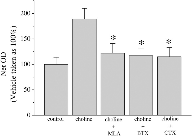
Fig 2. Choline effect on proliferation in MCF-7 cells.
Approximately 104 MCF-7 cells were seeded into microwell plates and grown for 3 days in the presence of vehicle (control), choline (10 mM), choline + methyllycaconitine (10 μM, MLA), choline + α-bungarotoxin (100 nM, BTX), and choline + α-Conotoxin RgIA (100 nM, CTX). Cells were harvested and growth was determined by an MTT assay. Bars represent means ± SEM of at least 3 independent determinations. Asterisk denotes significant difference from choline treated group with p < 0.05.

