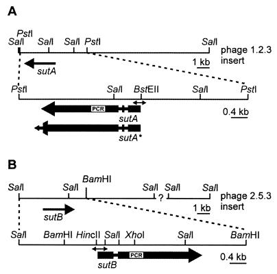
FIG. 1.
Physical map of the genomic DNA fragments containing the sutA (A) and sutB (B) genes. Genomic DNA fragments present in phages 1.2.3 and 2.5.3 carrying the sutA and sutB gene are depicted. The sutA and sutB open reading frames are indicated by the large, thick arrows, in which the narrow regions represent introns. For sutA, two versions are depicted, designated sutA and sutA*, the latter representing an extended open reading frame which would result if the last intron were spliced out. The sequences of the enlarged fragments (PstI-PstI for sutA and SalI-BamHI for sutB) are available in the GenBank database. The positions of the PCR fragments that were used to isolate the genes are indicated by the white boxes designated PCR. Regions that were used to probe expression in the Northern analysis are indicated by the double-headed arrows.

