
Figure 2.
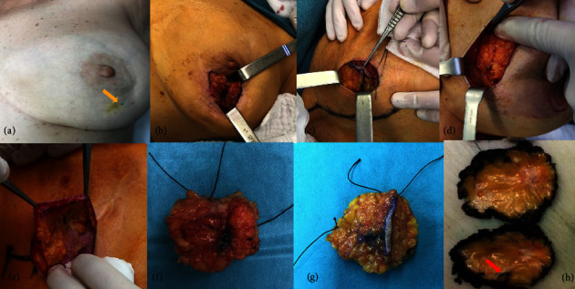
Surgical and pathological aspects of carbon localization in breast cancer tumors. During breast-conserving surgery, an incision is made over the carbon injection site (yellow arrow (a)) whenever possible. After carbon identification (b, c, e), the breast tumor lesion is excised with a macroscopic surgical margin (d). The breast surgical specimen is oriented (f, g) in a standardised way and sent to the pathology department for intraoperative gross pathological margin evaluation (h). The red arrow (h) shows the peritumoral carbon marking on the sliced surgical specimen.