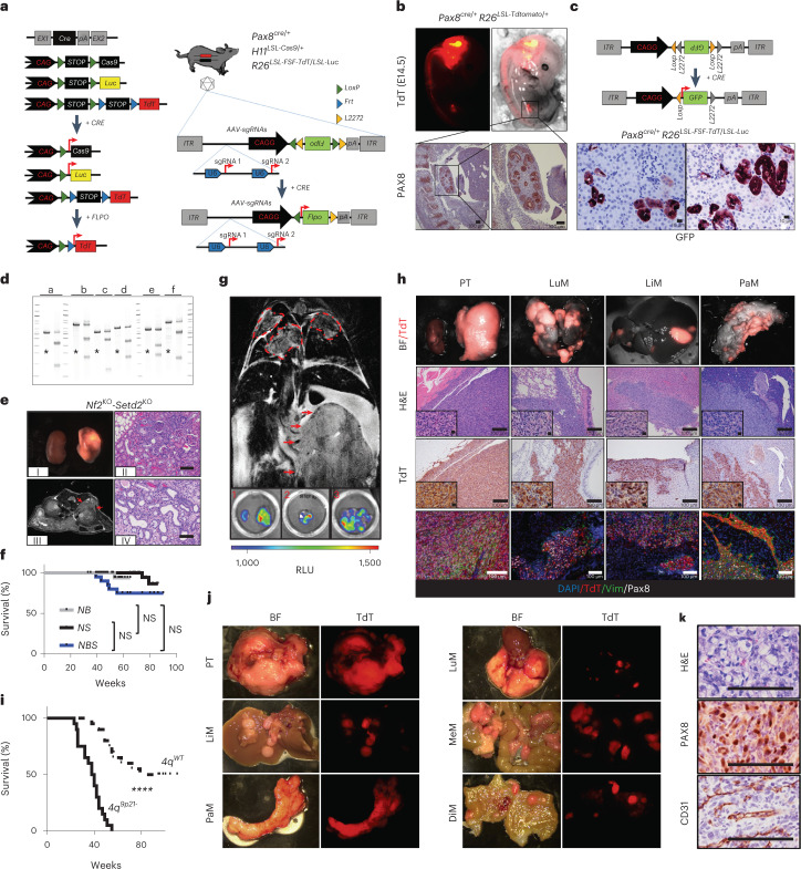
Fig. 1. SM-GEMM of RCC.
a, Schematic showing the SM-GEMM design. Cancer-specific loss-of-function mutations are introduced via intraparenchymal delivery of AAV particles carrying specific sgRNA combinations. b, Representative E14 Pax8Cre/+ -R26LSL-TdT/+ embryos. The activation of the fluorescent reporter TdT can be readily appreciated in the developing hindbrain, notochord and kidney. n = 5 embryos. c, Schematic showing the AAV-based tracing system carrying a FLEx-GFP-reported sequence. IHC analysis on representative FFPE sections stained with a GFP-specific antibody. n = 5 mice. d, T7-endonuclease assay validating sgRNA for Trp53 (a), Nf2 (b), Bap1 (c), Setd2 (d), Cdkn2a (e), Cdkn2b (f) and negative controls (*). Images representative of n = 3 independent experiments. e, Pathological characterization of murine RCC obtained through somatic mosaic knockout of Nf2 and Setd2. (I) Gross specimens collected 8 months posttransduction; (II) axial T2 MRI scan displaying a small cortical lesion 8 months posttransduction; and (III) and (IV) hematoxylin and eosin (H&E)-stained sections from well-differentiated tumors collected at 6 and 8 months posttransduction, respectively. f, Kaplan–Meier analysis of cancer-specific survival of mice affected by Nf2KO-driven tumors. NB: Nf2KO-Bap1KO (n = 40 mice); NS: Nf2KO-Setd2KO (n = 20 mice); NBS: Nf2KO-Setd2KO-Trp53KO (n = 24 mice). P = 0.23, 0.054, 0.12. g, Upper panel, representative coronal T2 MRI scan at 3 months posttransduction in Nf2KO-Setd2KO-4q9p21 mice. Red arrows, primary tumor mass; red dashed lines, lung metastasis. Bottom panels, representative luminescence scans of mouse organs. 1, primary tumor; 2, lung metastasis; 3, liver metastasis. Images representative of n = 2 experiments. h, Characterization of Nf2KO-driven murine tumors upon genetic targeting of the murine locus syntenic to human 9p21.3 (4q9p21): representative macroscopic images (top panels), H&E, IHC and IF analysis (lower panels). Images representative of n = 2 experiments. i, Kaplan–Meier analysis of cancer-specific survival of mice affected by VhlKO-driven tumors with (n = 20 mice) or without (n = 20 mice) 4q9p21 loss, P = 1.18 × 10⁻⁸. j,k, Characterization of VhlKO-driven murine tumors upon genetic targeting of 4q9p21 locus: representative macroscopic images (j), H&E and IHC analysis of specific clear cell RCC markers (PAX8 and CD31) are shown (k). PT, primary tumor; LuM, lung metastasis; LiM, liver metastasis; PaM, pancreatic metastasis; MeM, mesenteric metastasis; DiM, diaphragm metastasis. Images representative of n = 2 experiments. NS, not significant; ****P < 0.0001 by log-rank (Mantel–Cox) test. Scale bar, 200 μm. BF, brightfield; E, embryonic day; FFPE, formalin-fixed paraffin-embedded; IF, immunofluorescence; MRI, magnetic resonance imaging; RLU, renilla luciferase.

