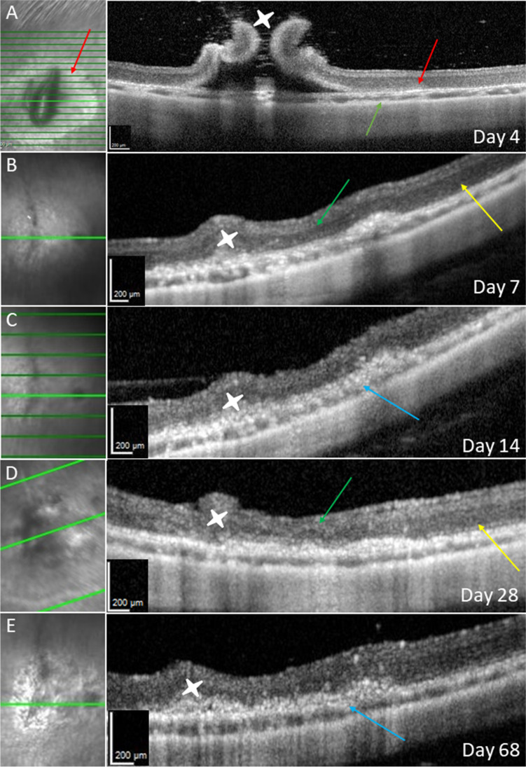
Fig. 2 .
OCT after RPE scraping. Day 4 (A): bRD with RPE wound, red arrows show RPE defect and corresponding outer retinal change. The orange arrow shows the choroid with almost normal thickness, without iatrogenic bleeding. Follow-up on day 7 (B), 14 (C), 28 (D), and 68 (E): green arrows showing retinal atrophy above the RPE wound, and blue arrows showing RPE hypertrophy. White stars show the retinotomy with initially high standing retinal margins. The yellow arrows show the surrounding tissue without RPE manipulation

