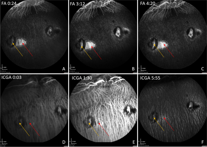
Fig. 4.
Representative FA/ICG with acquisition time (min:sec) at 1 week post-operatively. In the FA, a hyperfluorescence in the area of the induced RPE wound (red arrow) without leakage is shown over time (A–C). In the late phase of the ICGA, the RPE wounds appear hyperfluorescent with signal blockage corresponding to that seen in FA, suggesting it originates from hyperpigmentation of the formerly created RPE defect (D–F). The yellow arrow marks the retinotomy site

