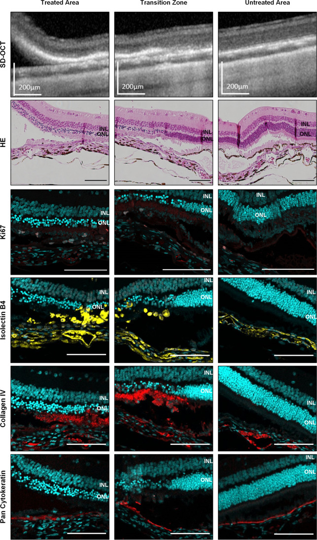
Fig. 5.
Visualization of the treated area, the transition zone, and untreated area as direct comparison by SD-OCT and histology at post-operative day 4. Hematoxylin and eosin (HE) staining already reveals atrophy of the photoreceptor outer segments and the outer nuclear layer (ONL) limited to the scraping site. The inner nuclear layer (INL) is not affected. Immunohistochemistry staining of the rabbit retina shows proliferating cells detected via Ki67 staining (red) which colocalize in part with subretinal microglia/macrophages shown by isolectin B4 staining (yellow). A strong deposition of collagen IV (red) is found around the scraping site beneath the ONL. Pan cytokeratin staining (red) for RPE already appears at the scraping site. Cell nuclei are stained with DAPI (cyan). Scale bar for SD-OCT = 200 µm, for histological images = 100 µm

