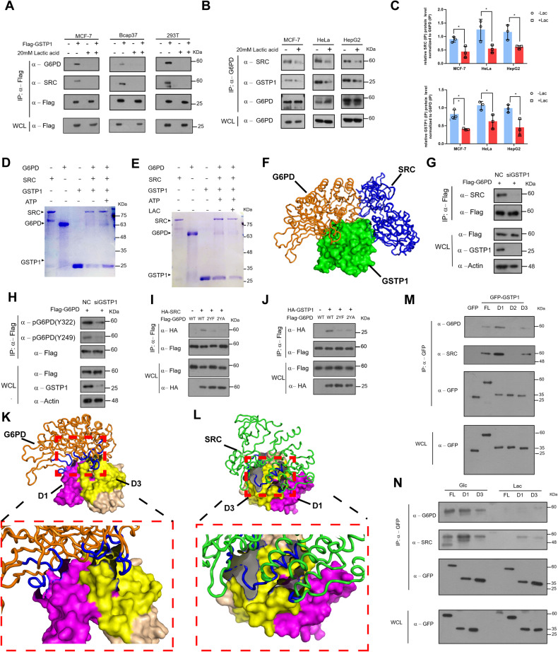
Fig. 3. A tripartite (GSTP1-G6PD-SRC) complex formation is modulated by lactate.
A Flag-tagged GSTP1 was overexpressed in cell lines (MCF-7, Bcap37, and 293 T) for 24 h. Cells were then incubated in glc medium (0.5 mM glucose) or lac medium (0.5 mM glucose with 20 mM lactic acid) for 12 h, and Flag-tagged GSTP1 protein immuno-purified for interaction detection. B MCF-7 cells/HeLa cells/HepG2 cells were incubated as in (A) for 12 h. Lysates were immuno-precipitated with anti-G6PD antibodies for endogenous interactions with GSTP1 and SRC. C Relative abundances of GSTP1 and SRC were obtained from 3 independent replicate experiments and normalized to immunoprecipitated G6PD endogenous protein in (B). *p < 0.05, **p < 0.01. D GST-SRC pull down to detect the GSTP1-G6PD-SRC interaction with or without ATP in vitro, as stained by CBB. E GST-SRC pull down to detect the GSTP1-G6PD-SRC interaction with or without lactic acid in vitro. F A modeled spatial arrangement of the GSTP1-SRC-G6PD complex. Green: GSTP1 (PDB ID: 3GUS); blue: SRC (PDB ID: 2H8H); Orange: G6PD(PDB ID: 2bhl). (G/H) Knockdown of GSTP1 decreases the interaction between G6PD and SRC (G) and overall G6PD phosphorylation level (H). I, J The interactions between GSTP1 (SRC) and G6PD decreased in G6PD mutants (2YF/2YA) in cells. HA-SRC (I), HA-GSTP1 (J), or HA-tagged empty control plasmids were co-transfected with WT (G6PD) or mutants (2YF/2YA) constructs for 24 h, and Flag-tagged G6PD was immunopurified and immunoblotted with indicated antibodies. K, L Modeled spatial arrangement of the GSTP1-SRC (L) or GSTP1-G6PD (K) complex. Orange: G6PD; Green: SRC; Pink: the 1-70AA region of GSTP1; Yellow: the 141-210AA region of GSTP1. M GFP, GFP-fused full length or indicated domains of GSTP1 were overexpressed in MCF-7 cells and immunoprecipitated by anti-GFP beads and examined through Western blots to check the interaction among G6PD, GSTP1 and SRC. N Proteins as indicated in (M) were overexpressed in MCF-7 cells, and cells treated with glucose-deprived (0.5 mM) media without or with 20 mM lactic acid to check the dynamic change of the tri-complex formation through Western Blots.

