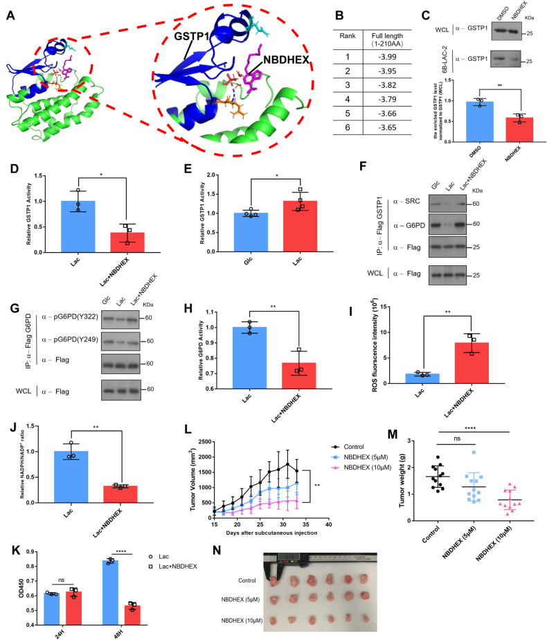
Fig. 5. A GSTP1-mediated lactic acid signaling promotes breast cancer cells proliferation.
A Modeled structure of parts of GSTP1 (PDB ID: 3GUS) with NBDHEX labelled in pink, the blue area represents parts of GSTP1 involved in NBDHEX binding. B Top 6 ranked binding energy values (kcal/mol) are shown. C GSTP1 treated with or without inhibitor NBDHEX was enriched via 6B-LAC-2 beads and examined through Western blots (Upper panel). The enriched GSTP1 via 6B-LAC-2 beads were determined (Lower panel). Data are presented as mean ± SD of triplicate experiments and analyzed by unpaired Students’ test. **p = 0.007. D The enzymatic activity of GSTP1 was examined with or without NBDHEX. *p = 0.0162. E The enzymatic activity of GSTP1 was examined with or without lactic acid in glucose-deprived cells. *p = 0.0446. F Flag-tagged GSTP1 was overexpressed in MCF-7 cell lines for 24 h. Cells were then incubated in glc medium (0.5 mM glucose) or lac medium (0.5 mM glucose with 20 mM lactic acid) or NBDHEX medium (0.5 mM glucose, 20 mM lactic acid and 10 µM NBDHEX) for 12 h, and Flag-tagged GSTP1 protein immuno-purified for interaction assays. G Flag-tagged G6PD was overexpressed in MCF-7 cell lines for 24 h. Cells were then incubated in glc medium (0.5 mM glucose) or lac medium (0.5 mM glucose with 20 mM lactic acid) or NBDHEX medium (0.5 mM glucose, 20 mM lactic acid and 10 µM NBDHEX) for 12 h, and the phosphorylation status was determined. H MCF-7 cells overexpressing flag-tagged G6PD were pretreated with or without 10 µM NBDHEX for 1 h, switched to 20 mM lactic acid and 10 µM NBDHEX for 12 h and enzymatic activity of G6PD measured. Data are presented as mean ± SD of triplicate experiments and analyzed by unpaired Students’ test. **p = 0.0096. I, J After treatments as in (D), the ROS levels (I) and NADPH/NADP+ ratio (J) were measured and analyzed by unpaired Students’ test. **p (ROS) = 0.0049; **p (NADPH/NADP+ ratio) = 0.0017. K MCF-7 cells were seeded in the 96-well plate, treated with or without 10 µM NBDHEX in media containing 20 mM lactic acid for indicated days, and measured at OD 450 nm by using CCK-8 kit. Data are presented as mean ± SD of triplicate experiments and analyzed by multiple t test. ****p (48H Lac vs 48H Lac NBDHEX) = 0.000049. L Tumor sizes were measured from day 15. Data are presented as mean ± SD of triplicate experiments and analyzed by unpaired Students’ test. **p (Control vs 10µM NBDHEX) = 0.0021. M Tumors were weighted at day 34. Data are presented as mean ± SD of triplicate experiments and analyzed by unpaired Students’ test. ****p (control vs 10µM NBDHEX) < 0.0001. N Pictures illustrating breast tumor burdens in nude mice grafted with MCF-7 cells and subjected to no or NBDHEX treatment.

