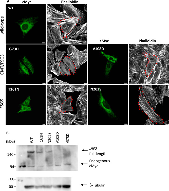
Figure 7.
Expression of INF2 variants causing either single FSGS or dual CMT-FSGS phenotypes in HeLa cells (A) Subcellular localization of INF2 variants and actin network. cMyc-tagged wild-type INF2 and pathogenic variants were transfected into HeLa cells. At 8 h post-transfection, the cells were fixed and stained with anti-cMyc antibody (green) and phalloidin (white). Two variants (G73D, V108D) cause a dual CMT/FSGS phenotype, whereas the other two (T161N, N202S) lead to FSGS alone. The number of actin stress filaments was markedly reduced in cells expressing the G73D, V108D variant compared to those expressing the T161N, N202S variant. Dashed lines highlight the cell outline. Bars = 10 μm. (B) Western blot analysis of INF2 variants. INF2 protein expression in HeLa cells expressing cMyc-tagged wild-type INF2 or the G73D, V108D or T161N and N202S variants were analyzed by western blot. FSGS variant proteins (T161N, N202S) were less abundant than wild-type protein. CMT/FSGS variants (V108D, G73D) were barely detectable, suggesting that this variant may be more vulnerable to degradation. After the stripping step, internal control of β-tubulin was re-probed on the identical membrane with the same exposure time as the INF2 detection (5 min). Original blots/gels are presented in Supplementary Figure S14.

