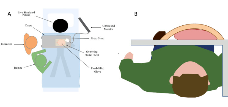
Figure 1. Original illustration of ultrasound pericardiocentesis model.
(A) Aerial view and (B) transverse view. The protective Mayo stand (gray) was placed over a live supine volunteer. Above the tray, a fluid-filled glove was surrounded by a plastic sheet. The fluid was aspirated from the glove under ultrasonographic guidance.
Image credits: Authors' original

