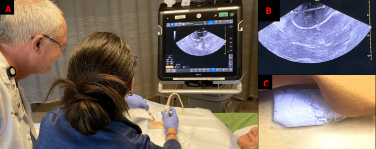
Figure 2. Photographs of the ultrasound pericardiocentesis model.
(A) A surgical educator guiding a trainee through ultrasound-guided pericardiocentesis with hybrid simulation setup using a volunteer mock patient. (B) Displayed ultrasound image from the simulator design. (C) Simulator construction with skin and drape reflected revealing the “pericardial fluid collection”.

