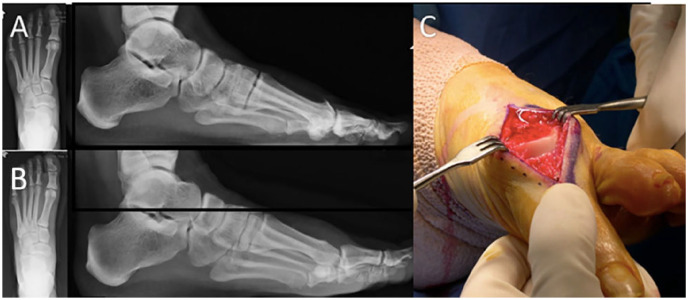
Figure 2.
Cheilectomy. (A) Radiographic images demonstrating well-preserved joint space with large dorsal osteophyte. (B) Radiographic image following cheilectomy. (C) Operative image following dorsal cheilectomy. Note the obvious debridement of osteophytes, removal of the dorsal articular surface and maintained joint alignment.

