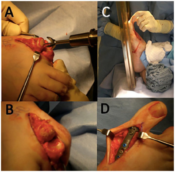
Figure 6.

Intraoperative photograph of first metatarsophalangeal joint arthrodesis: (A) Conical reamers used to debride residual cartilage and create congruent surfaces for fusion. (B) The metatarsal head following joint preparation. Note the drill holes used to perforate the subchondral bone surface. (C) Flat plate used to judge great toe clearance in the proposed position of fusion. (D) Dorsal plate in place. Not seen is the lag screw placed from distal to proximal that compresses the joint surface.
