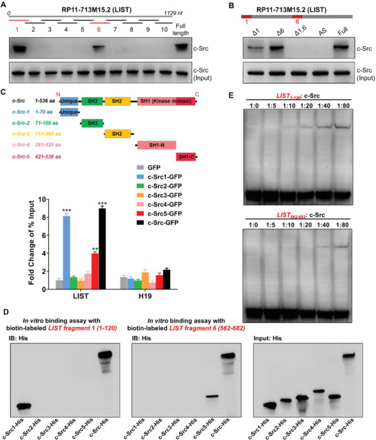
Figure 2.

Direct interaction between LIST and c‐Src. A) Interactions between fragmented LIST and c‐Src. Biotin‐labeled LIST fragments (≈120 nt each) and full‐length LIST were used for protein pull‐down in cell line 5637, followed by western blot analysis of c‐Src. B) Interactions between LIST truncation and c‐Src. biotin‐labeled LIST truncations (that lack one or both of fragments 1 and 6) and full‐length LIST were used for protein pull‐down in cell line 5637, followed by western blot analysis of c‐Src. C) Different c‐Src truncations were fused with GFP and expressed in cell line 5637. Enrichment of LIST was detected using GFP‐RIP‐qPCR. H19 was used as a negative control. The error bars represent the SD of three replicates. D) The biotin‐labeled LIST fragment (1 or 6) was incubated with Dynabeads (MyOne Streptavidin C1), and then mix with the purified His‐tagged c‐Src truncations and full‐length proteins in binding buffer, followed by western blotting for His. E) EMSA image of a biotin‐labeled LIST fragment (1 or 6) binding to different concentration gradients of c‐Src proteins. Two fmol of the labeled LIST fragment were used for each EMSA reaction.
