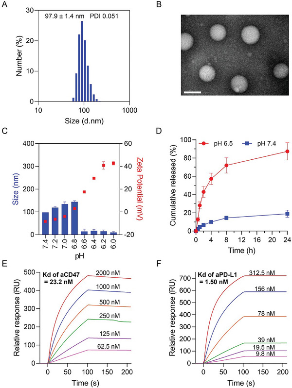
Figure 1.

Characterization of NCPA. A) The hydrodynamic diameter of NCPA. B) TEM images of NCPA in pH 7.4. C) Hydrodynamic diameter changes of NCPA in different pH buffers. D) The protein release profiles of NCPA in different pH buffers. E) The in vitro SPR analysis of CD47 antibody binding to CD47 protein. F) The in vitro SPR analysis of PD‐L1 antibody binding to PD‐L1 protein.
