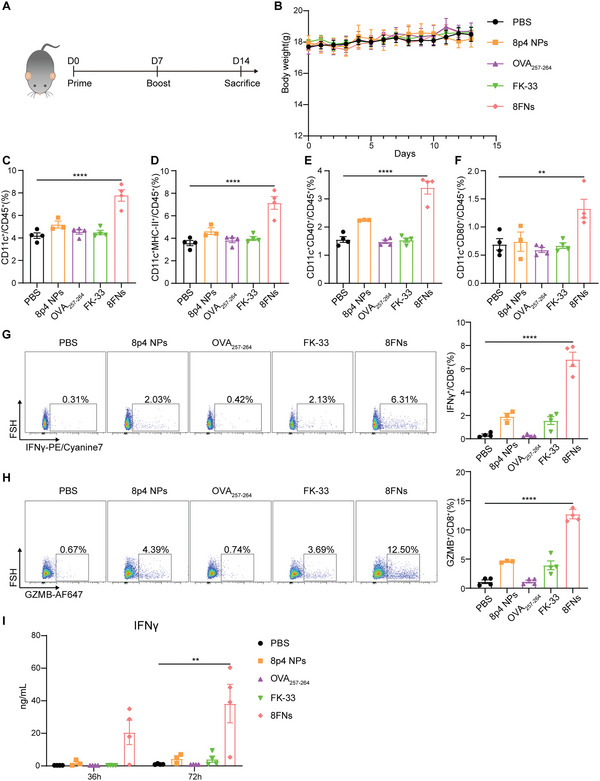
Figure 3.

8FNs activate DCs in lymph nodes and elicit specific CD8+ T cell response safely in vivo. A) Mice were immunized subcutaneously with PBS (n = 4), 8p4 NPs (n = 3), peptide OVA257‐264 (n = 4), peptide FK‐33 (n = 4) or 8FNs (n = 4) on day 0, day 7 and sacrificed on day 14 for subsequent analysis. B) The mice body weights of the indicated groups for 14 days. C–F) Flow cytometry analysis results of the percentage of CD11c+, CD11c+MHC‐II+, CD11c+CD40+, and CD11c+CD80+ DCs in lymph nodes. G,H) Flow cytometry analysis results of the percentage of IFNγ +CD8+ T cells (G) and the percentage of GZMB+CD8+ T cells (H) in splenocytes stimulated by peptide OVA257‐264 on 72 h. I) The splenocytes were isolated from pretreated mice and stimulated with peptide OVA257‐264 (10 µg mL−1) in a medium. The IFNγ production was measured by ELISA at 36 and 72 h. (B,C–I) Datadisplayed as the mean ± SEM. Significance (**P < 0.01, ****P < 0.0001) in (C–I) was estimated by one‐way ANOVA with Dunnett's multiple comparisons test.
