
Figure 7.
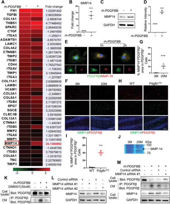
MMP14 mediates PDGF‐BB‐induced PDGFRβ shedding. A) Gene‐array analysis on rh‐PDGFBB‐treated or control human brain pericytes for 3 days. Heatmaps showed the magnitude of differential expression with fold change (on the right) of each gene. B) Quantitative real‐time PCR analysis of MMP14 mRNA expression in human brain pericyte with or without rh‐PDGFBB treatment for 24 h. n = 3. C) Western blot analysis of MMP14 protein expression in primary human brain pericyte with or without rh‐PDGFBB treatment for 24 h. D) Quantification of the relative intensity of MMP14 using Image J. n = 3. E) Primary human brain pericytes were treated with rh‐PDGFBB for 1 or 2 h. Double‐immunofluorescence staining of the cells was performed using antibodies against MMP14 (red) and PDGFRβ (green). F,H) Representative confocal images of PDGFRβ (red) and MMP14 (green) double‐immunofluorescence staining in DG region of 3‐ and 22‐month‐old WT mice (F) and 6‐month‐old PdgfbcTG mice and WT littermates (H). DAPI stains nuclei as blue. Scale bar, 100 µm. G,I) Quantification of the percentage of MMP14+ cells in PDGFRβ + pericytes in hippocampus. n = 5. J) Representative images of gel zymography showing the activity of MMP in the hippocampus of 3‐ and 20‐month‐old WT mice. K) Primary human brain pericytes were treated with rh‐PDGFBB in the presence or absence of MMP inhibitor GM6001 for 24 h, and culture medium (CM) was collected. PDGFRβ expression in cell lysate was detected by Western blot analysis (upper panel), and the cleaved sPDGFRβ in CM was immunoprecipitated by a specific PDGFRβ antibody followed by Western blot analysis of PDGFRβ expression (lower panel). L) Two different MMP14 siRNAs or scrambled control siRNA were individually transfected into human brain pericytes. MMP14 (upper panel) and GAPDH (lower panel) expression levels were detected by Western blot analysis. M) Primary human brain pericytes were transfected by MMP14 siRNA or scrambled control siRNA followed by rh‐PDGFBB treatment for 24 h. PDGFRβ expression in cell lysate was detected by Western blot analysis (1st Row), and the cleaved sPDGFRβ in CM was immunoprecipitated by a specific PDGFRβ antibody followed by Western blot analysis of PDGFRβ expression (2nd Row). MMP14 (3rd Row) and GAPDH (4th Row) expression levels in cell lysate were also detected by Western blot analysis. Data are shown as the mean ± SD, ***p<0.001, unpaired two‐tailed Student's t test.