
Figure 8.
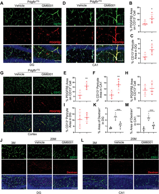
GM6001 rescue MMP14‐induced PDGFRβ shedding and aged‐associated BBB leakage. A–I) PdgfbcTG mice were treated with GM6001 or vehicle every other day by i.p. injection for 2 weeks. Representative confocal images of PDGFRβ (red) and CD13 (green) double‐immunofluorescence staining in DG (A), CA1 (D), and cortex (G). DAPI stains nuclei as blue. Scale bar, 100 µm. Quantification of the percentage of PDGFRβ expression area on CD13+ cells (B, E, H) and CD13+ pericyte area (C, F, I) in the three regions. J–M) 18‐month‐old mice were treated with GM6001 or vehicle every other day by i.p. injection for 4 weeks. Representative confocal images of CD31 (green) and Dextran (red) double‐immunofluorescence staining in DG (J) and CA1 (L). DAPI stains nuclei as blue. Scale bar, 100 µm. Quantification of the percentage of Dextran+ signal area in these two areas (K and M). n = 5. Data are shown as the mean ± SD, *p<0.05, **p<0.01, ***p<0.001, as determined by unpaired two‐tailed Student's t test (for two group comparison) or One‐way ANOVA (for multiple group comparison).