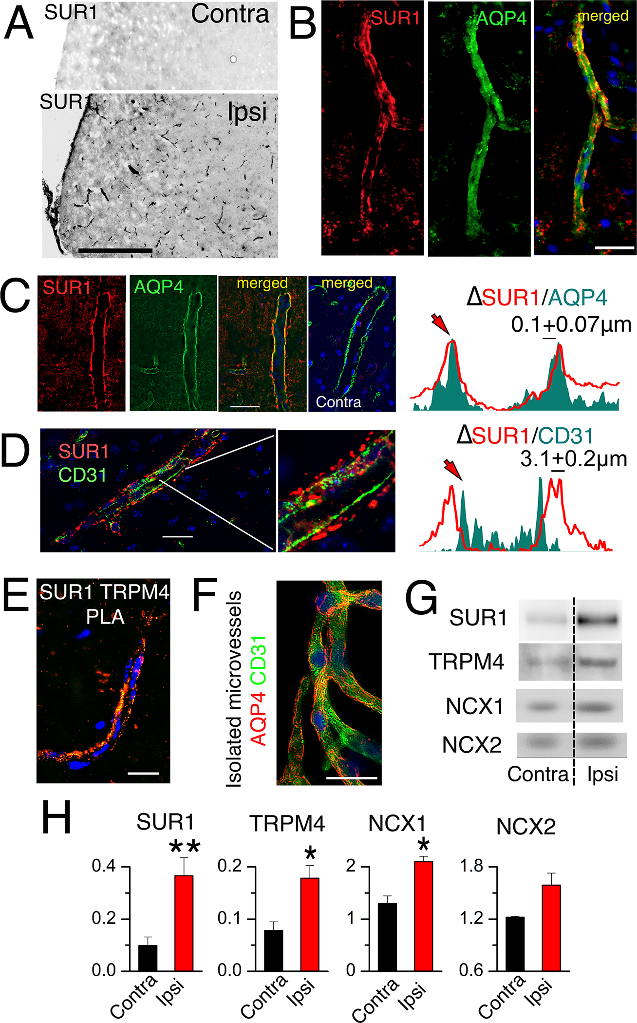
Fig. 1. SUR1-TRPM4 and NCX1 are expressed in post-ischemic astrocyte endfeet.

(A) Immunohistochemical analysis of expression of SUR1 (black) in ipsilateral (Ipsi) and contralateral (Contra) microvessels in brain tissue sections from mice after MCAO for 2 hours followed by reperfusion for 24 hours. Scale bar, 200 μm. (B to D) As described in (A) but with 6-hour reperfusion, immunohistochemistry for expression of SUR1 (red) in AQP4+ astrocyte endfeet (green; merged image) in microvessels within ipsilateral (B,C) and contralateral tissues (C), and for SUR1 (red) and CD31 (endothelium marker; green) in ipsilateral tissues (D). Nuclei are stained blue with DAPI (4′,6-diamidino-2-phenylindole). Right, fluorescent intensity profiles for the respective pairs. Profiles are representative and data are mean ± S.E. from N = 30 (C) and 24 (D) measurements in ipsilateral tissues from 3 mice. Scale bars, 25 μm. (E) PLA imaging for SUR1-TRPM4 heteromers lining a microvessel in ipsilateral tissues isolated from mice as described in (B–D). Image is representative of 5 mice. Scale bar, 25 μm. (F) Immunolabelling for CD31 (green) and AQP4 (red) in isolated microvessels from ipsilateral tissues from mice as described in (B–D). Image is representative of 4 mice. Scale bar, 25 μm. (G and H) In mice as described in (B–D), microvessels isolated from ipsilateral (Ipsi) and contralateral (Contra) tissues were studied by immunoblot for SUR1, TRPM4, NCX1, and NCX2 (G). Data (H) are mean ± S.E. from 3 or 4 mice per group, normalized to β-actin. *P<0.05 and **P<0.01 by t-test.
