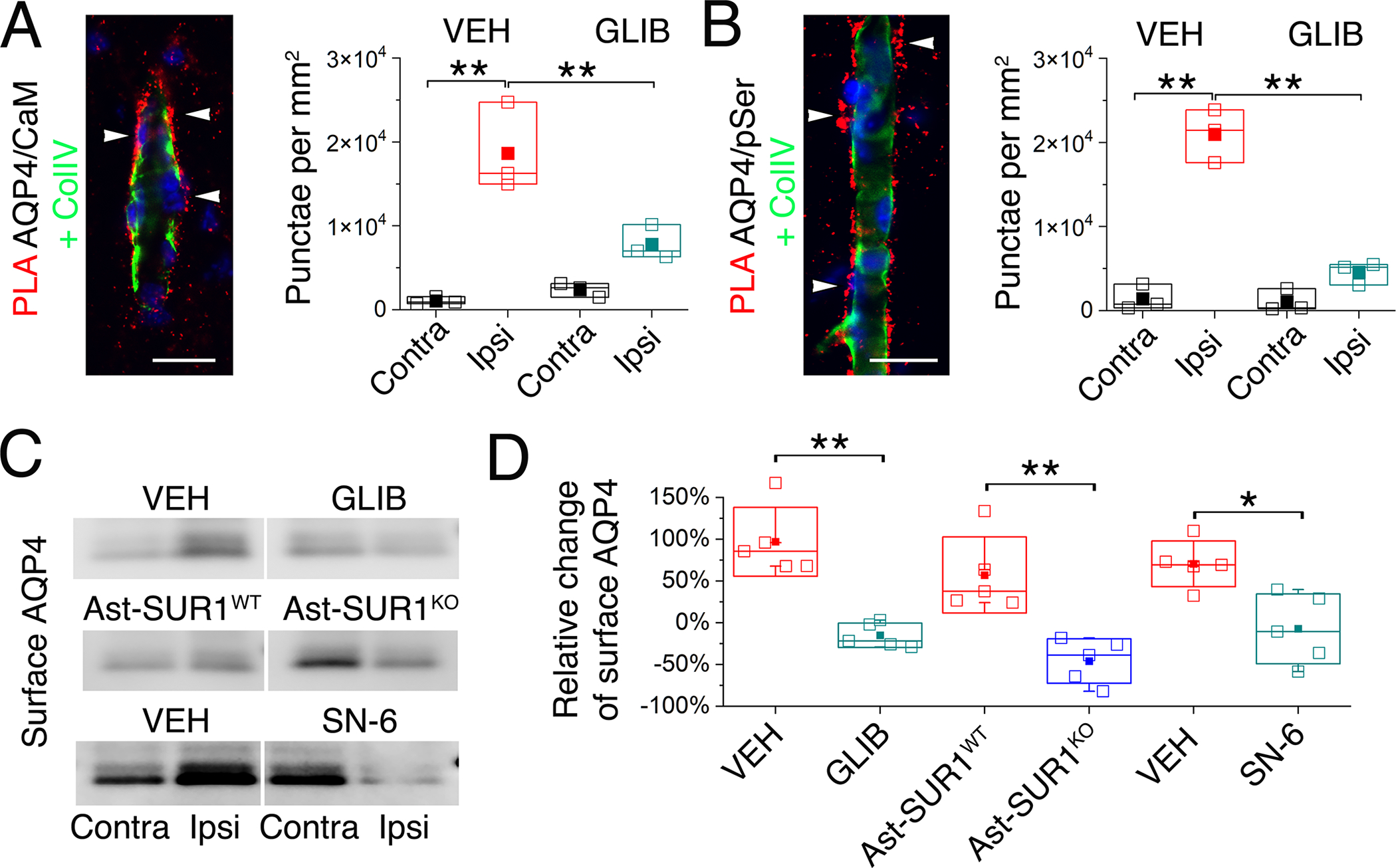
Fig. 3. Cell membrane localization of aquaporin-4 increases after ischemia and requires SUR1-TRPM4 and NCX1.

(A and B) Proximity ligation assay (PLA) (red) showing co-association of AQP4 with calmodulin (CaM) (A), and serine phosphorylation (pSer) of AQP4 (B), both located outside of the microvascular collagen IV layer (ColIV; green) following MCAO/R (2 hours MCAO with 6 hours reperfusion). The number of PLA punctae (arrowheads) per mm2, mean ± S.E. from 3 mice per group, is plotted for ipsilateral (Ipsi) and contralateral (Contra) hemispheres from mice administered vehicle (VEH) or glibenclamide (GLIB) at reperfusion. Nuclei stained blue with DAPI **P<0.01 for contra vs. ipsi; ##P<0.01 for VEH vs. GLIB, both by ANOVA. Scale bars: 25 μm. (C and D) In mice as described in (A,B), and additionally WT mice administered SN-6, and in a mouse with astrocyte-specific deletion of Abcc8/SUR1 (Ast-SUR1KO) and a littermate control (Ast-SUR1WT), immunoblot was performed for ipsilateral (Ipsi) and contralateral (Contra) cell-membrane AQP4 (C), with densitometric data for cell-membrane AQP4 (ipsilateral relative to contralateral) quantified (mean ± S.E.) for the 6 conditions (D); *, P<0.05; **, P<0.01 by ANOVA; 5 mice/group.
