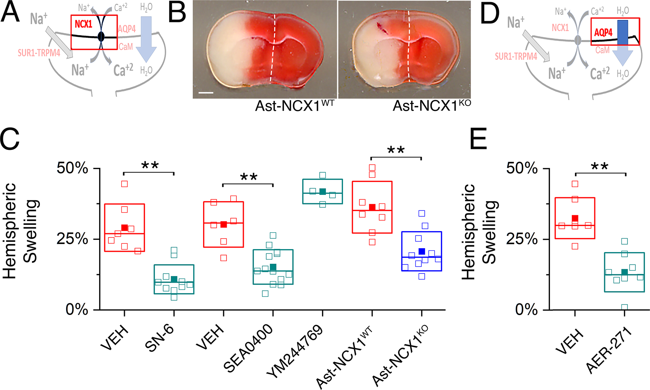
Fig. 6. NCX1 and AQP4 play critical roles in brain swelling independent of infarct size.

(A) Proposed model of the functional axis comprised of SUR1-TRPM4, NCX1 and AQP4, with emphasis on NCX1. (B) Images of TTC-stained coronal brain sections following MCAO/R in a mouse with deletion of Slc8a1/NCX1 in astrocytes (Ast-NCX1KO) and a littermate control (Ast-NCX1WT). Scale bar: 1 mm. Ischemia was induced by MCAO for 2 hours followed by reperfusion for 24 hours. Images are representative of 8 mice per group. (C) In mice as described in (B) with infarct volumes >40 mm3, ipsilateral hemispheric swelling following MCAO/R was quantified in slices from WT mice administered vehicle (VEH) or SN-6 or SEA0400 or YM244769 at reperfusion, and mice with conditional astrocyte-specific deletion of Slc8a1/NCX1 (Ast-NCX1KO) and littermate controls (Ast-NCX1WT). Data are mean ± S.E. from 4 to 13 mice per group. **P<0.01 by ANOVA. (D) Proposed model of the functional axis comprised of SUR1-TRPM4, NCX1 and AQP4, with emphasis on AQP4. (E) In mice as described in (B) with infarct volumes >40 mm3, ipsilateral hemispheric swelling following MCAO/R was quantified in slices from WT mice administered vehicle (VEH) or AER-271 at reperfusion. Data are mean ± S.E. from N = 7 or 8 mice per group. **P<0.01 by ANOVA.
