
Fig. 8.
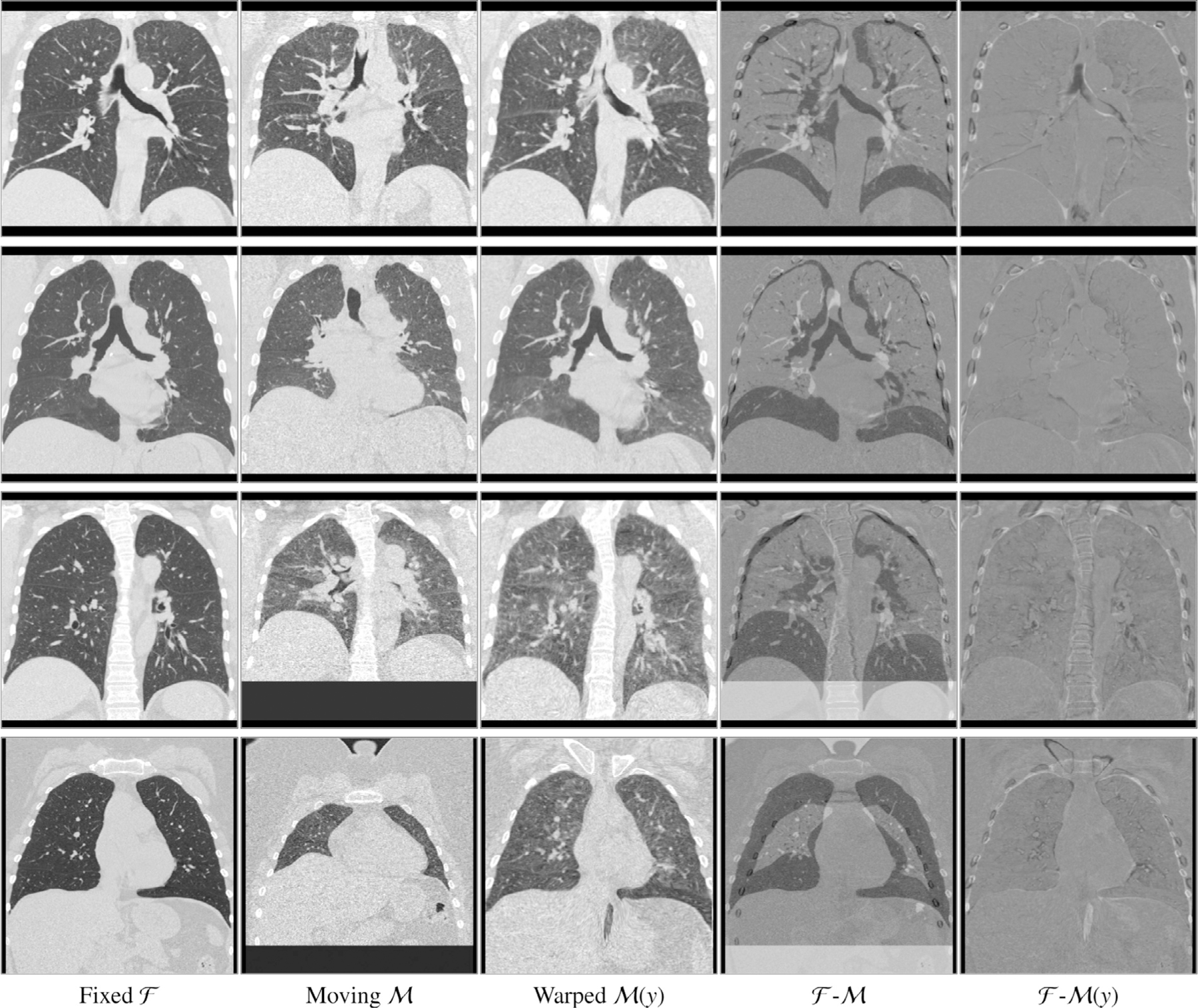
Example coronal slices extracted from four exemplary cases. Input images and , the warped moving image , the difference image (fourth column) and the difference image after registration with the proposed method (fifth column). In all cases the respiratory motion was successfully recovered and most inner structures are well aligned. Due to altered density of lung tissue during breathing, intensity changes occur and therefore higher values in the difference images are reached without registration errors.