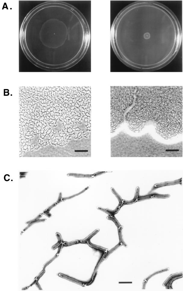
FIG. 2.
Macroscopic and microscopic analysis of mc2155 spreading on the surface of agarose plates. (A) Halo formation on 0.3% (left) and 0.8% (right) agarose plates containing M63 salts with no added source of carbon. Plates were inoculated by poking a single colony from a 0.2% glucose M63 agar plate and transferring it to the center of the plate. The photograph was taken after 5 days of incubation at 37°C. (B) Phase-contrast images of the edges of the spreading halos shown in panel A. Bar, 25 μm. (C) Electron micrograph of cells spreading on a 0.3% agarose–M63 salts plate. A Formvar carbon-coated grid was placed directly over the spreading halo and cells were stained with 1% uranyl acetate. Bar, 2 μm.

