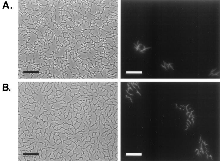
FIG. 3.
Growth accompanies mycobacterial spreading. A 1:100 mix of GFP-labeled (light) and unlabeled (dark) mc2155 cells grown as described in Materials and Methods were plated on the surface of 0.3% M63 salts– (A) and 7H9 (with no added carbon source)– (B) agarose plates. Photographs were taken after 2 days of incubation at 37°C. Phase-contrast images showing the continuous spreading halo are on the left, and fluorescent micrographs of the same fields showing the locations of GFP-labeled cells are on the right. Bars, 25 μm.

