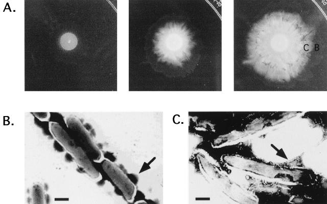
FIG. 5.
Pattern formation in a spreading Sm-1 colony. A 25-μl aliquot of a saturated Sm-1 culture was plated onto a 7H9–ADC–0.3% agarose plate. (A) Pictures of the spreading colony taken 1, 2, and 3 days after inoculation. (B and C) Electron micrographs of cells taken at day 3 from the transparent periphery (B) and opaque interior (C) of a spreading colony. Cells were negatively stained with 2% phosphotungstic acid. Bar, 1 μm. Arrows mark the structures discussed in the text.

