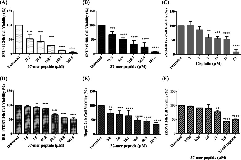
Fig. 3.
37-mer peptide dose-dependent cytotoxicity. A SNU449 displayed a significant decrease in cell viability (approx. 50%) upon treatment with peptide (71.2 μM) at 24 h. B Treatment of SNU449 for 48 h with peptide showed a similar trend to 24 h treatment. Cells also experienced significant cell death (approx. 60% viability) at 71.2 μM exposure. C SNU449 cells were screened with 48 h treatment of Cisplatin. 7 μM of cisplatin treated SNU449 showed similar cell viability to peptide treatment (71.2 μM). D 1BR-hTERT cells were treated with the peptide for 24 h. Cells showed similar viability (approx. 50%) at a lower treatment concentration (60.8 μM) compared to SNU449 cells (71.2 μM). E HepG2 peptide treatment at 24 h displayed similar viability (approx. 50%) at a lower concentration (30.4 μM) compared to more advanced hepatocellular carcinoma SNU449 cells (71.2 μM). F SKOV3 treated with peptide for 24 h showed a decrease in cell viability (approx. 80%) at 24 μM. Treatment with 33 nM of Cisplatin reduced cell viability by approx. 50%. SKOV3 displayed greater sensitivity to the peptide, as cytotoxicity started to decrease from 0.24 μM of treatment. (** P < 0.01, *** P < 0.001, **** P < 0.0001, n = 3)

