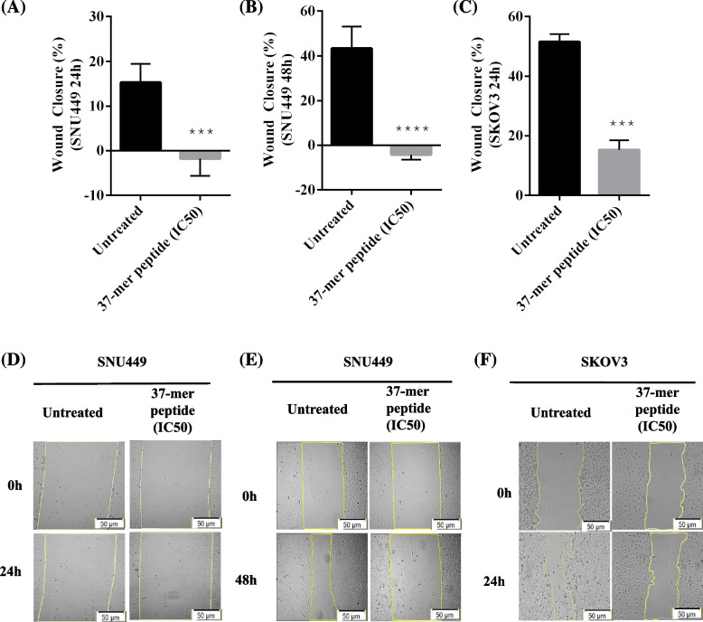
Fig. 6.
37-mer peptide treatment hindered SNU449 and SKOV3 cell migration. Highly confluent cells were subject to vertical scratches to observe cell migration effect when treated with IC50 of the peptide. A and B Percentage wound closure of SNU449 cells measured at 24 h and 48 h time points at the IC50 concentration respectively. Wound closure decreased by approximately 20% between untreated and treated (76.4 μM) cells, in addition, the closure also diminished by approximately 50% following 48 h exposure (88.4 μM). C Average wound closure of untreated SKOV3s and 37-mer peptide treated (27.5 μM) cells. Treated SKOV3 closure was significantly reduced, by up to 40%, compared to untreated cells (*** P < 0.001, **** P < 0.0001, n = 3). D Representative wound closure images of SNU449 cells treated with peptide for 24 h. E Similarly, shows wound healing images of SNU449 cells treated for 48 h. F Wound closure of SKOV3 cells untreated and treated with peptide. Cell images are processed at magnification scale of 50 μm

