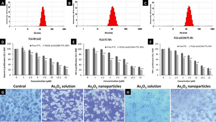
Fig. 5.
Nanoparticle delivery system of AML treatment Note: (A-C). Size distribution of three kinds of nanoparticles including void PLGA nanoparticles (PLGA-NPs), PLGA nanoparticles encapsulating parthenolide (PLGA-PTL-NPs), PLGA nanoparticles with anti-CD44 and encapsulating parthenolide (PLGA-antiCD44-PTL-NPs). Cell proliferation after treated with free PTL and PLGA-antiCD44-PTL-NPs against (D) Kasumi-1, (E) KG-1a and (F) THP-1 cell lines. Cell proliferation is expressed as mean ± S.E.M (n = 3). One-way ANOVA was used followed by the Newman-Keuls post-test (*P < 0.05, **P < 0.01 compared to PTL). (Figure source from the reference [59], color figure can be viewed at mdpi.com). Optical inverted microscopy images of NB4 cells incubated with As2O3 solution or its nanoparticles for 48 h (magnification, ×40) at the concentrations of 1.5 µmol/L (G) and 3.0 µmol/L (H). (Figure source from the reference [53], color figure can be viewed at spandidos-publications.com)

