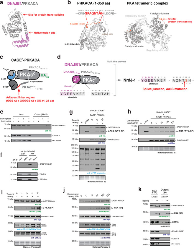
Extended Data Fig. 8 |. Design of DNAJB1-CAGEN and CAGEC-PRKACA for inducible kinase activation in cells.
a, Crystal structure of the DNAJB1/PRKACA fusion oncoprotein (PDB: 4WB7). The native gene fusion site and the potential ligation site for protein splicing are indicated. b, Left – crystal structure of mouse PRKACA (PDB: 4DFX) highlighting the myristate tail bound to the allosteric site in the catalytic domain. A flexible linker colored in red is a potential site for insertion of the CAGEC module. Right – crystal structure of PKA tetrameric complex (PDB: 3TNP). The location of the CAGEC module insertion site is indicated in red. c, Schematic depicting the design of the CAGEC-PRKACA construct. The N-terminal alpha-helix of PRKACA containing the N-myristoylation site (1–37 aa) is connected to the N-terminus of the CAGEC module via a 24 amino acids flexible linker. d, Schematic of the split site used within DNAJB1/PRKACA incorporation of the CAGE modules. A38 amino acid site in the PRKACA is mutated to serine in the N-terminus of split PRKACA for protein trans-splicing reaction by NrdJ-1Cage. e, f, Immunoblots of the HA-tagged CAGEC-PRKACA construct expressed in HEK293T cells cultured with YnMyr and enriched using the workflow described in fig. S7F. CAGEC-PRKACA construct was enriched following YnMyr metabolic labeling and biotin conjugation (e). By contrast, the G2A mutation of CAGEC-PRKACA failed to be enriched in the workflow (f). g, Proximity-induced CPS in HEK293T cells co-expressing DNJAB1-CAGEN (Myc-tag) and CAGEC-PRKACA (HA-tag). Cells were treated with DMSO or rapalog (100 nM) for indicated time-points before immunoblotting. X refers to untransfected cells, whereas J-PKA refers to cells expressing the positive control DNAJB1/PRKACA fusion (HA tagged). h, Similar to panel (g) but showing the dose-response to rapalog at the 24-hour timepoint. i, Proximity-induced CPS in AML12 cells co-expressing DNJAB1-CAGEN (Myc-tag) and CAGEC-PRKACA (HA-tag). Cells were treated with DMSO or rapalog (100 nM) for indicated time-points before immunoblotting. j, Similar to panel (i) but showing the dose-response to rapalog at the 6-hour timepoint. k, Immunoblots of HSP70 co-immunoprecipitated with HA-tagged splice product. A pulldown assay was performed using the lysates from stable AML12 cells treated with DMSO or rapalog (100 nM) for 24 hours. Data in (e-k) are representative of n=3 independent experiments. CAGEN refers to ‘NrdJ-1NCage-FKBP,’ and CAGEC refers to ‘PKAcauto-FRB-NrdJ-1CCage’ in (e-k).

