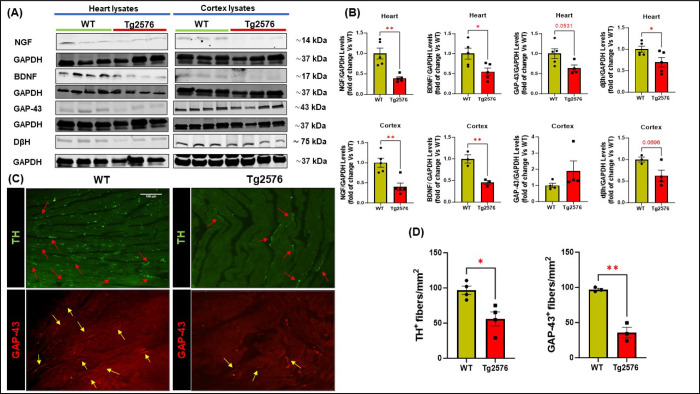
Figure 5: The neuro-signaling pathway is compromised in brain and heart of Tg2576 AD mice.
(A-B): Representative immunoblots (A) and densitometric quantitative analysis (B) showing levels of NGF, BDNF, GAP-43, and DβH, in total cardiac (top panels) and cerebral cortex (bottom panels) lysates from WT and Tg2576 mice. (C-D): digital images (C, scale bar 100μm) and quantifications (D) showing cardiac adrenergic nerve fibers, labeled with anti-tyrosine-hydroxylase (TH, in green), and cardiac regenerating nerve endings, labeled with anti-neuronal regeneration marker (GAP-43, in red) in heart sections from WT and Tg2576 mice. n=3– 5 mice/group. Data are presented as a mean±SEM. *P<0.05, **P<0.01 and vs WT. Student t-tests have been performed between the 2 groups.

