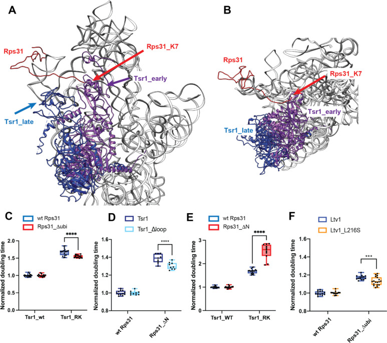
Figure 8: Rps31 binding requires Tsr1 movement to the beak.
(A) Composite structure of pre-40S ribosomes (6FAI) with Tsr1 in the early (6FAI) and late position (6WDR) and showing Rps31 from mature ribosomes (3JAM). 6FAI and 3JAM ribosomes were overlaid by matching Rps18, and 6WDR was overlaid onto these by matching 18S rRNA. (B) Structural detail of a top view of the structure in panel A highlighting the N-terminus of Rps31 near Tsr1 in the early position. Note that residues 1–6 are not resolved, the first residue visible is K7. (C) Doubling times (normalized to wt Tsr1) for yeast cells expressing either wt Rps31 or Rps31 lacking the N-terminal ubiquitin fusion (Rps31Δubi), and either wt Tsr1, or Tsr1_RK. (D) Doubling times (normalized to wt Rps31) for yeast cells expressing either wt Rps31 or Rps31 lacking the N-terminal extension (Rps31ΔN), and either wt Tsr1, or Tsr1_Δloop. (E) Doubling times (normalized to wt Tsr1) for yeast cells expressing either wt Rps31 or Rps31 lacking the N-terminal extension (Rps31ΔN) and either wt Tsr1, or Tsr1_RK. (F) Doubling times (normalized to wt Rps31) for yeast cells expressing either wt Rps31 or Rps31Δubi, and either wt Ltv1, or Ltv1_L216S. Significance was tested using an unpaired t-test. ****, P<0.0001.

