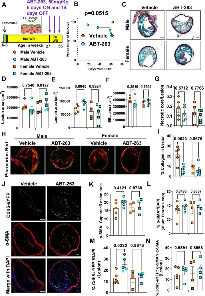
Figure 4. 263 Treatment of Apoe−/− mice with advanced lesions with a reduced dose (50mg/kg/bw) of ABT- decreased collagen content within BCA lesions and was also associated with increased mortality.
(A) Experimental design, EC-lineage tracing Apoe−/− mice were fed a WD for 18 weeks followed by 50mg/kg/bw ABT-263 treatment on WD for 9 weeks. (B) Probability of survival (Kaplan-Meier curve). (C) Representative 10x images with 100μm scale bar of MOVAT staining of the BCA. (D) Lesion area from C. (E) Lumen area from C. (F) External elastic lamina (EEL) area from C, for outward remodeling. (G) Necrotic core area normalized to lesion size. (H) Representative 10x images with 100μm scale bar of Picrosirius red staining on Brachiocephalic Artery (BCA). (I) Quantification of mature (red) collagen content normalized to lesion area from figure H. (J) Representative confocal images of co-staining for eYFP (for detecting EC), α-SMA+, and DAPI in advanced BCA lesions. (K) α-SMA+ cap area normalized to lesion area (α-SMA+ cap area/Lesion area). (L) Quantification of the percentage α-SMA+ (α-SMA+/DAPI) cells in the fibrous cap. (M) Quantification of the percentage EC-derived (Cdh5-eYFP+/DAPI+ cells in the lesion, and (N) quantification of the percentage EC-derived α-SMA+ (Cdh5-eYFP+ α-SMA+/α-SMA+) cells in lesions. The two-way ANOVA method was used for statistical analysis in D, G, and K-L, and biologically independent animals are indicated as individual dots. Error bars show the SEM. A Mantel-Cox test used for statistical analysis in B. The p-values are indicated on the respective graphs.

