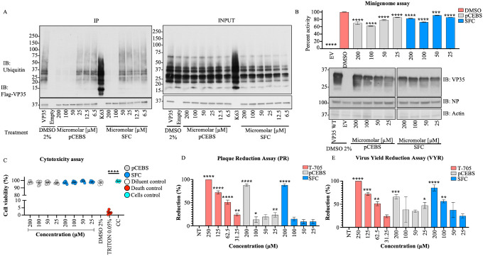
Figure 7. pCEBS and SFC compounds inhibit interactions between VP35 and K63-linked polyubiquitin chains and correlate with loss of viral polymerase activity and virus replication.
A) Flag-VP35 bound to anti-Flag beads were incubated for 1h at room temperature with different concentrations of pCEBS or SFC, followed by incubation with recombinant purified unanchored K63-linked polyUb chains (2–7). VP35-Ub complexes were eluted with Flag-peptide and analyzed by Immunoblot. B) 293T cells were transfected with minigenome components and 4 hours post-transfection cells were treated with pCEBS and SFC compounds at different concentrations. 50 hours later cells were lysed for luciferase assay. C) Cytotoxicity test (CyQUANT MTT Cell Viability Assay ThermoFhisher) using pCEBS and SFC at different dilutions D) Plaque reduction and (E) Virus Yield Reduction assays, the cells were infected by 1 hour and after 1h the treatment was made with pcEBS, SFB compound or DMSO: Dimethyl sulfoxide with the overlay. The number of plaques in each set of compound dilution were converted to a percentage relative to the untreated virus control. The percent of activity from the ratio of luciferase and renilla (Luc/ren) was calculated. Data are depicted as Mean + SEM of the two independent assays in triplicate. Tukey’s multiple comparisons tests. p < 0.001 **, p < 0.0001 ***, p < 0.00001 ****.

