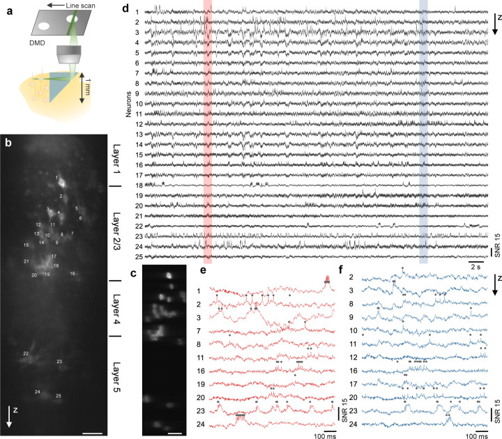
Figure 5. Side-on voltage imaging across multiple cortical layers with an implanted microprism.
(a) Schematic illustration of side-on cortical imaging with an implanted microprism.
(b) Side-on confocal image of Voltron2 fluorescence, demonstrating simultaneous view of neurons across cortical layers 1–5. Scale bar, 50 µm.
(c) Averaged Voltron2 fluorescence image with 25 neurons targeted within the FOV. Scale bar, 50 µm.
(d) Voltron2 fluorescence traces for the 25 targeted neurons shown in (b) over a continuous 45 s recording.
(e,f) Zoomed-in fluorescence traces of active neurons in the shaded regions shown in (d).

