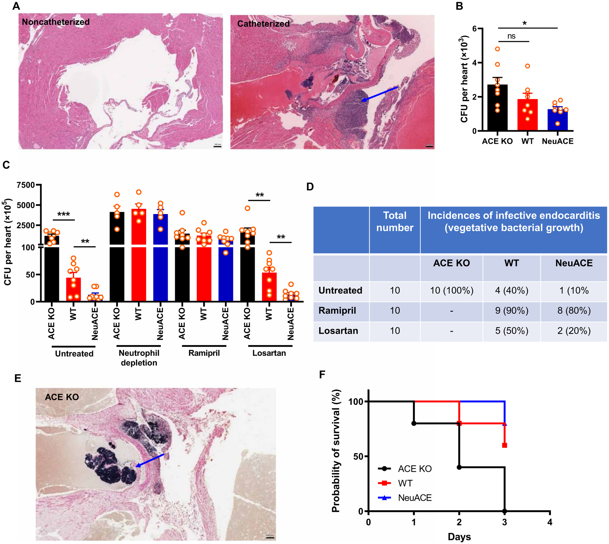
Fig. 3. Inhibition of ACE increased the incidence of infective endocarditis in MRSA-infected mice.

(A) Representative images of hematoxylin and eosin–stained murine heart transverse sections with or without aortic valve damage by catheter insertion are shown. The blue arrow indicates aortic valve damage and infiltration of cells. Scale bars, 100 μm. (B) Bacteria were counted per heart in ACE KO, WT, and NeuACE mice challenged with intravenous MRSA (about 1 × 10⁷ CFU/100 μl) without valve damage. (C) Bacteria were counted per heart in mice challenged with intravenous MRSA with aortic valve injury. ACE KO, WT, and NeuACE mice were untreated, depleted of neutrophils, or treated with ramipril or losartan. n ≥ 7. (D) Incidence of infective endocarditis in catheterized ACE KO, WT, and NeuACE mice as indicated by Gram staining (n = 10 per group). (E) A representative image of Gram staining shows bacterial accumulation in the heart of a catheterized ACE KO mouse challenged with intravenous MRSA. The blue arrow shows vegetative bacterial growth. Scale bar, 100 μm. (F) Survival of ACE KO, WT, and NeuACE mice after aortic valve damage and MRSA challenge is shown (n = 5 per group). A one-way (B) or two-way ANOVA (C) with Bonferroni’s correction for multiple comparisons was used to analyze group comparisons, and data are presented as means ± SEM. *P < 0.05, **P < 0.01, and ***P < 0.001.
