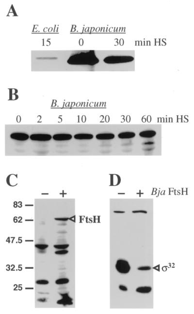
FIG. 4.
Immunodetection of FtsH protein in E. coli and B. japonicum extracts. E. coli C600 or B. japonicum 110spc4 cells were grown to mid-exponential phase at 30°C. After a reference sample had been taken, the culture was shifted to 43°C and samples were collected at the indicated time points after heat shock (HS). Crude cell extracts were prepared, separated on a sodium dodecyl sulfate–12% polyacrylamide gel, and subjected to Western blot analysis using anti-E. coli FtsH serum (U. Jenal, Basel, Switzerland; original source, P. Bouloc, Paris, France; 1,000-fold dilution). Only the area around 70 kDa is shown. Panels A and B represent two independent experiments with different sampling periods. A 10-fold-diluted E. coli extract was loaded in panel A in order to avoid a strong signal due to the use of a homologous antiserum. For the experiments shown in panels C and D, E. coli AR3291/pBAD18-Cm and AR3291/pRJ5188 were grown at 30°C in LB medium with arabinose (0.1% [wt/vol]) to mid-exponential phase and samples were subjected to Western blot analyses. The absence (−) or presence (+) of B. japonicum (Bja) FtsH expressed from pRJ5188 is indicated. Bands representing B. japonicum FtsH in panel C and E. coli ς32 in panel D are labeled. The sigma factor was detected by the use of anti-E. coli ς32 serum (B. Bukau, Freiburg, Germany; 3,000-fold dilution). The molecular masses of marker proteins are indicated in kilodaltons on the left.

