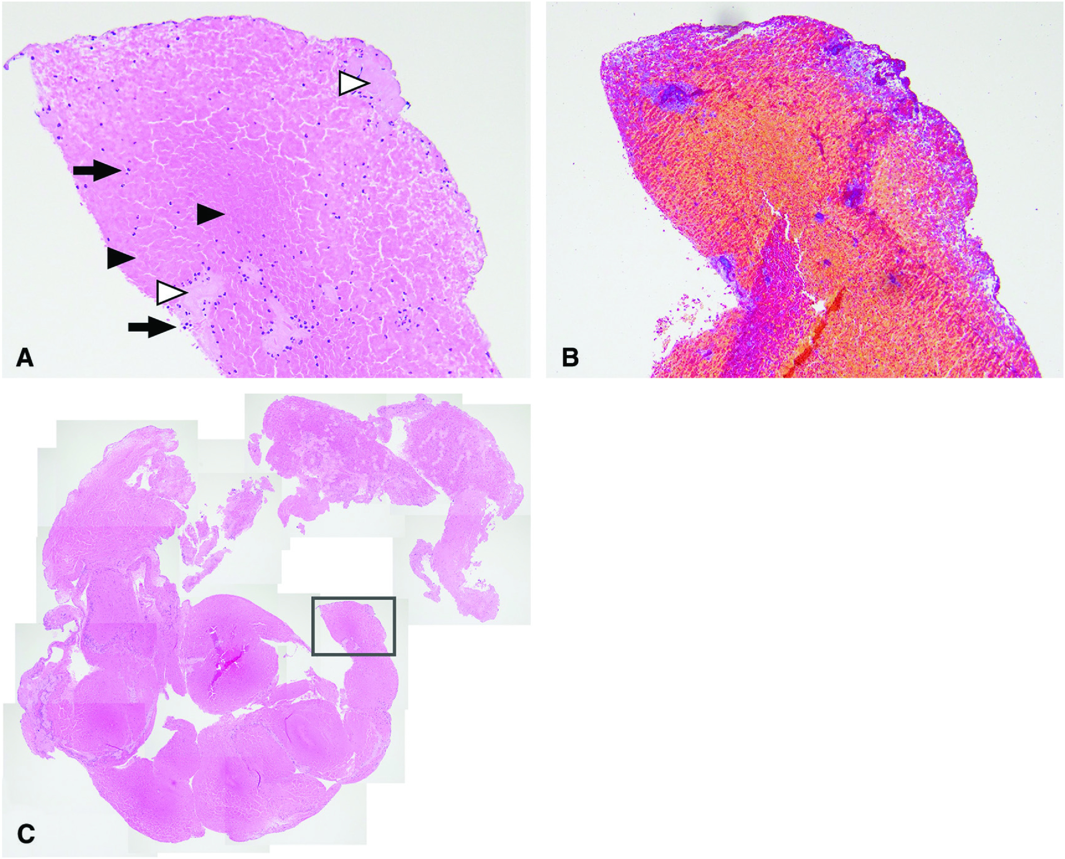
Fig. 1. Microscopic view of the thrombi retrieved in case 4. (A) Hematoxylin–eosin stained section (100× magnification), WBC (arrows), RBC (arrowheads), and F/P (white arrowheads); (B) Masson’s trichrome-stained section (100× magnification); (C) hematoxylin-eosin stained section (20× magnification). F/P: fibrin/platelet; RBC: red blood cell; WBC: white blood cell.

