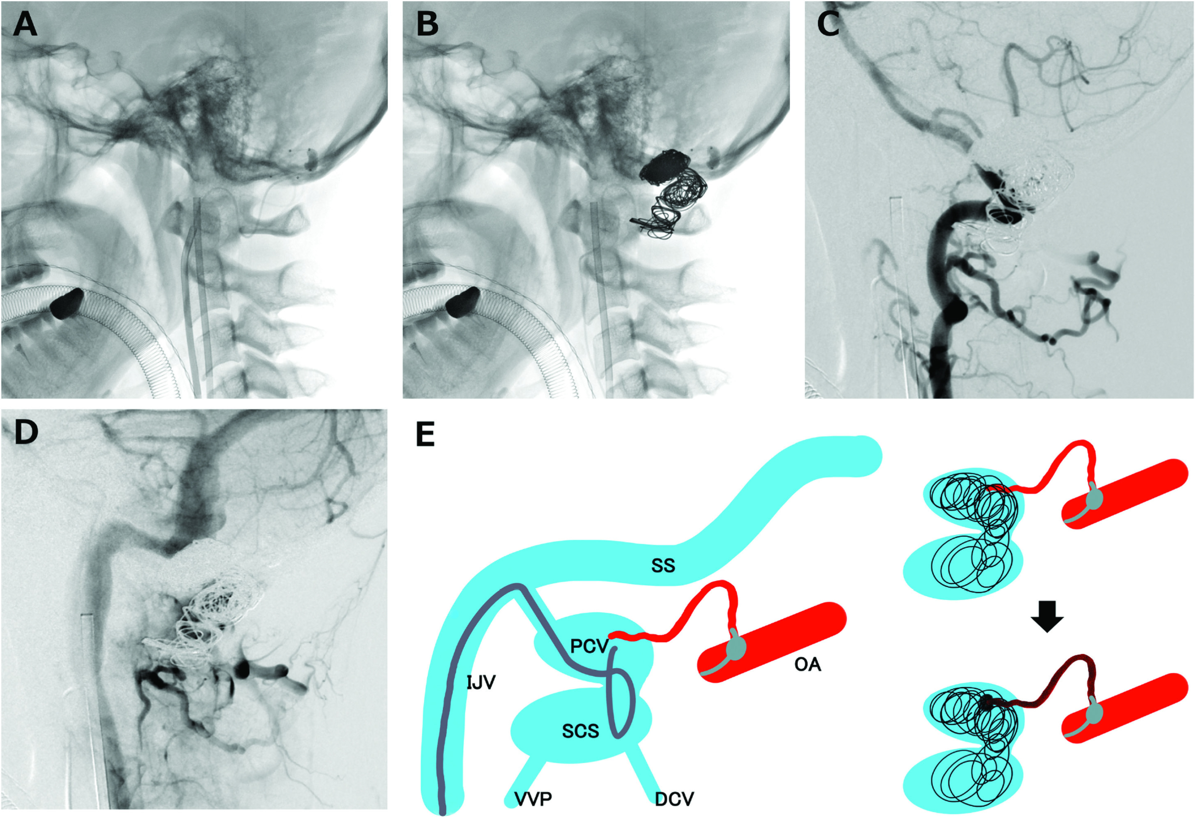
Fig. 3. (A) Microcatheter is navigated from the internal jugular vein to the PCV, making a loop at the SCS to embolize the SCS in addition to the PCV packing. A balloon catheter is advanced into the dural branch of the right occipital artery for flow control. (B) Platinum coils are placed at the venous pouch and part of the SCS to prevent onyx migration into distal vessels. (C) High-flow shunt is decreased in the right VA angiogram. (D) Coil protrusion to the internal jugular vein is denied. (E) Schema of the strategy in our case. Transvenous coil embolization of the PCV and part of the SCS was performed, following the Onyx injection from the meningeal branch of the right occipital artery. DCV: deep cervical vein; IJV: internal jugular vein; OA: occipital artery; PCV: posterior condylar vein; SCS: suboccipital cavernous sinus; SS: sigmoid sinus; VA: vertebral artery; VVP: vertebral venous plexus.

