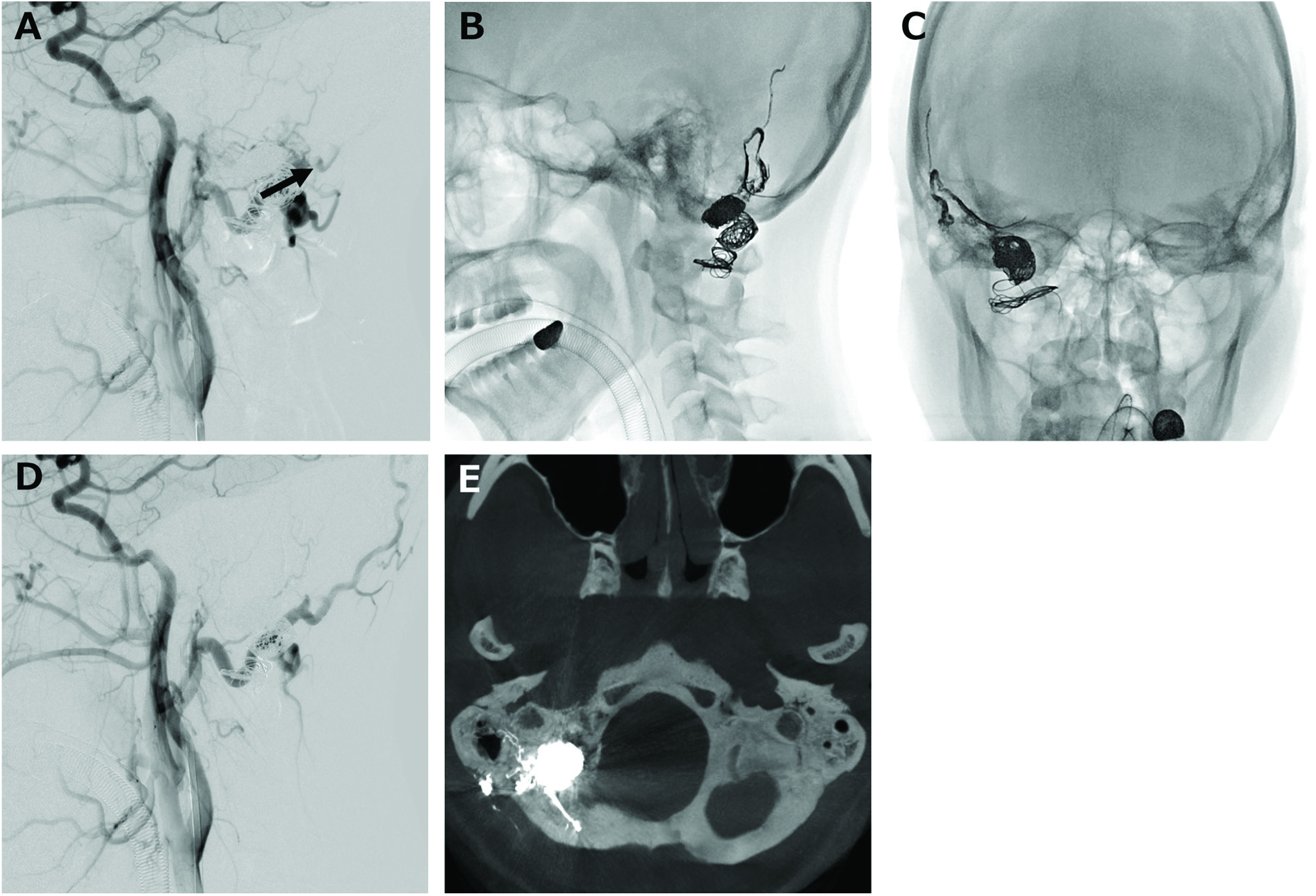
Fig. 4. (A) Lateral view of the common carotid arteriogram shows the feeders from the meningeal branch of the right occipital artery remaining after the coil embolization. Frontal (B) and lateral (C) views of the craniogram. Onyx was injected from the inflated balloon catheter placed at the dural branch of the occipital artery. (D) Lateral view of the common carotid arteriogram shows the DAVF being completely disappeared. (E) CT demonstration of the onyx cast filling the occipital artery and posterior meningeal artery. DAVF: dural arteriovenous fistula.

