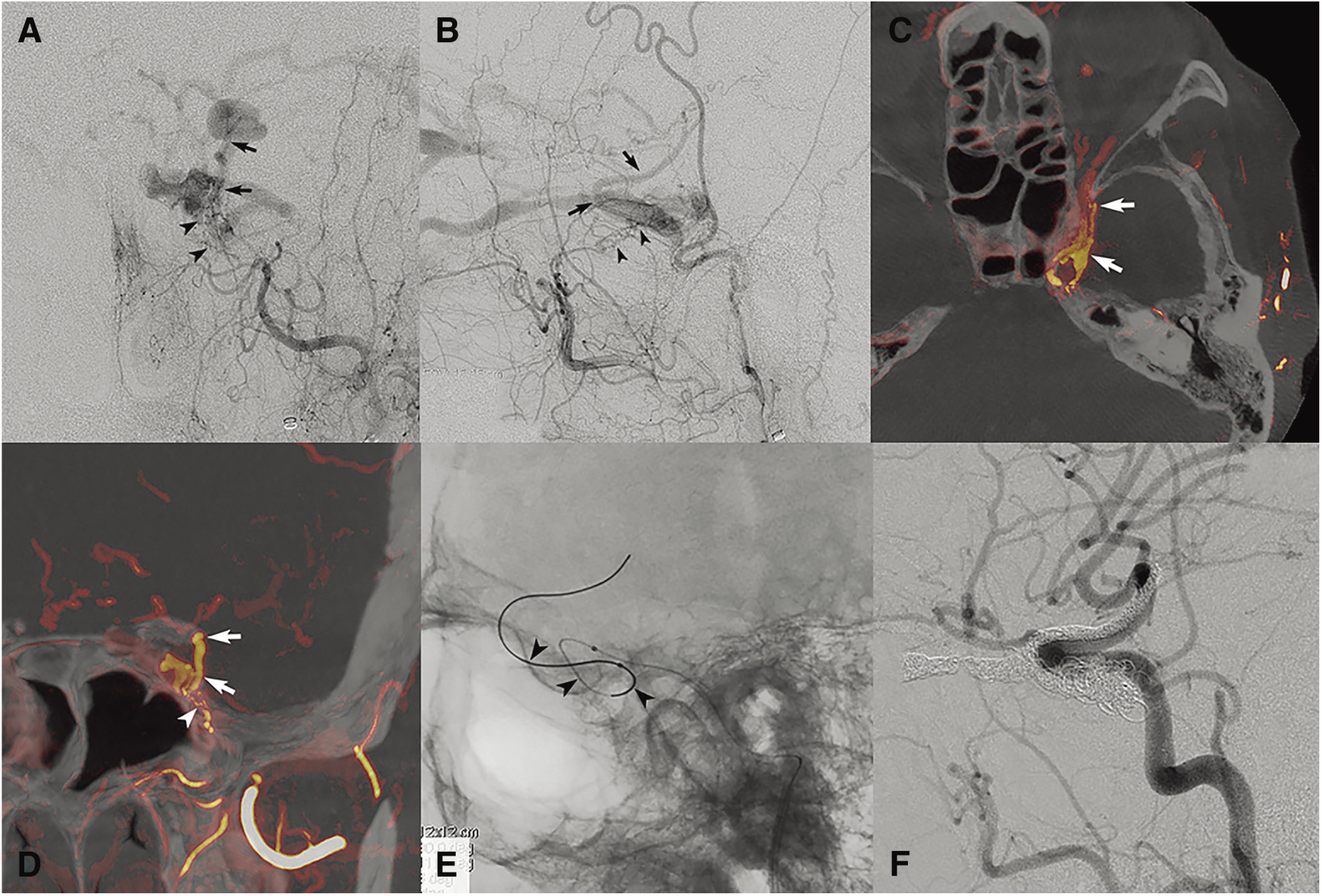
Fig. 11. A case with left CS dural arteriovenous fistula involving laterocavernous sinus. (A and B) Left external carotid arteriogram (A, frontal view; B, lateral view) shows cavernous dural arteriovenous fistula mainly fed by artery of foramen rotundum (arrowheads) and draining into inferior ophthalmic vein and SMCV (arrows). (C and D) Color fusion images of 3D external carotid arteriogram (C, axial reconstruction; D, coronal reconstruction) demonstrate draining into SMCV via laterocavernous sinus (arrows). Laterocavernous sinus is connected with CS at posterior compartment. Note arteriovenous shunt at the caudal part of laterocaverous sinus (D, arrowhead). (E) Fluorogram after navigation of microcatheter and guidewire (arrowheads) into laterocavernous sinus and SMCV through connection at posterior segment. (F) Lateral view of left common carotid arteriogram after coil packing of SMCV, ophthalmic vein, laterocavernous sinus, and CS shows complete disappearance of dural arteriovenous fistula. CS: cavernous sinus; SMCV: superficial middle cerebral vein.

