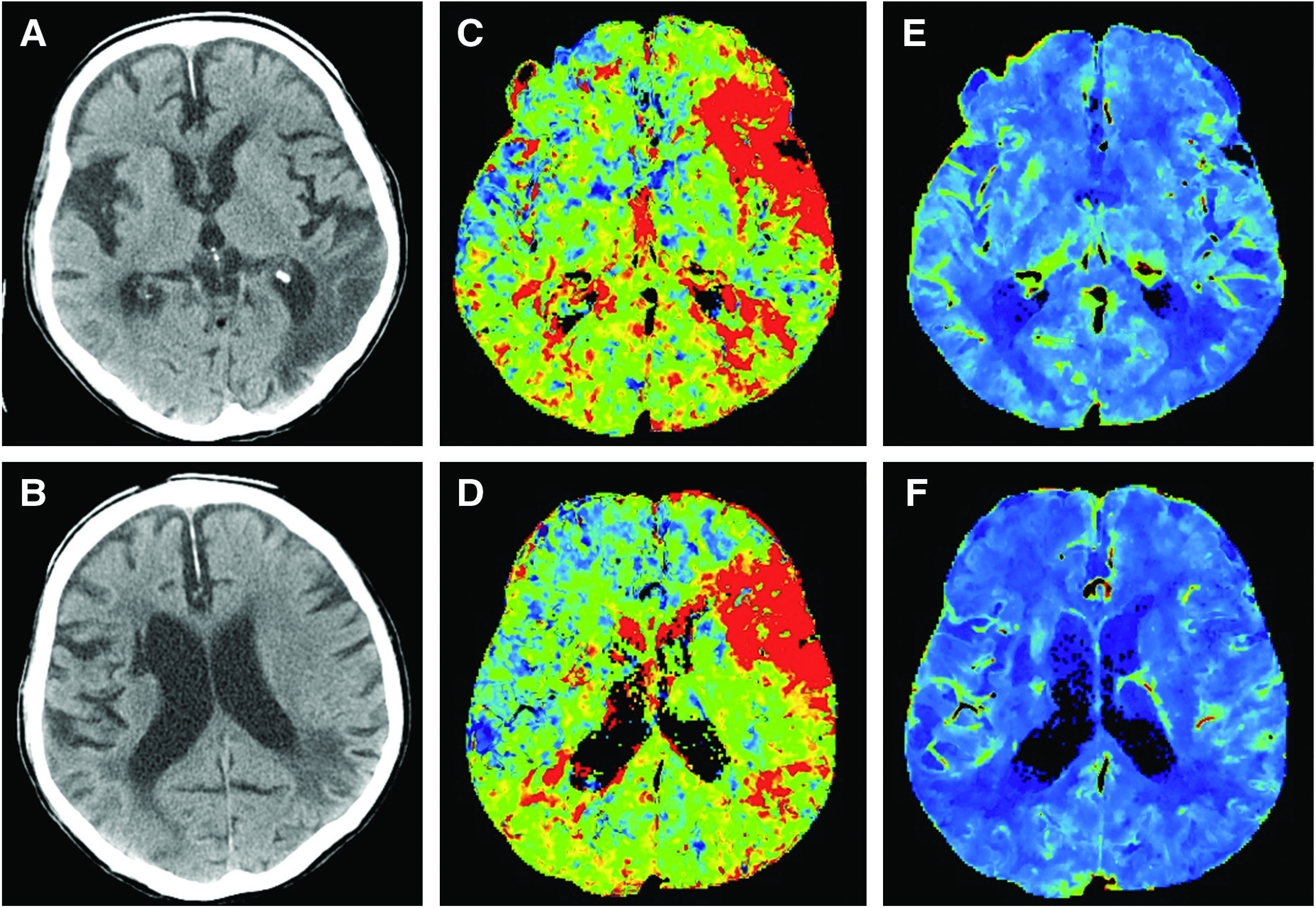
Fig. 1 . On plain CT (A and B), old cerebral infarction was present in the left parietotemporal lobe. On CT perfusion, delayed Tmax (C and D) was noted in the left middle cerebral artery territory but no decrease in CBV (E and F) was noted in the same area. CBV: cerebral blood volume; Tmax: time-to-maximum of residue function.

