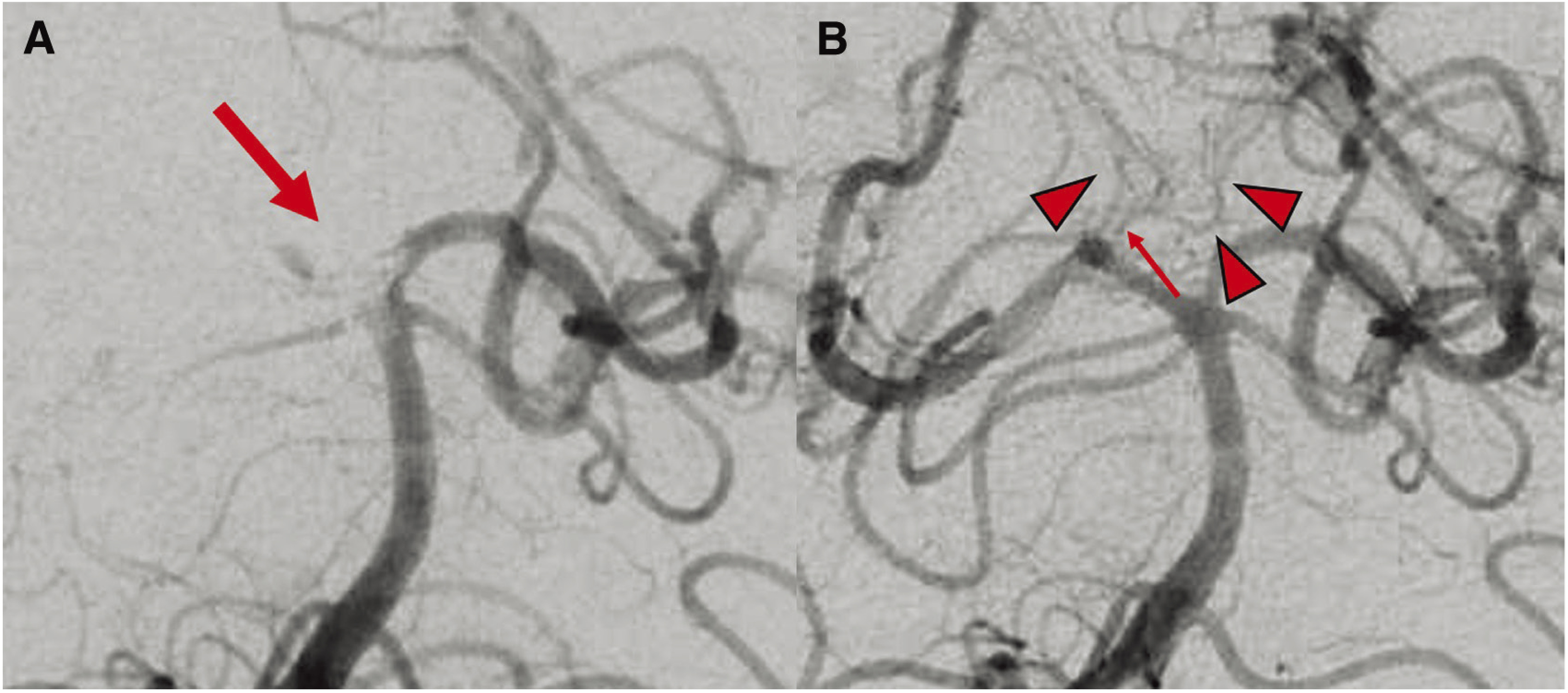
Fig. 3. DSA of the right vertebral artery. (A) DSA shows the thrombus from the top of the BA to the right P1 segment (arrow). (B) Postoperative DSA shows the perforator branch (arrow) from the right P1 segment, which is the common trunk for the bilateral PTA (arrowheads). BA: basilar artery; PTA: paramedian thalamic arteries.

