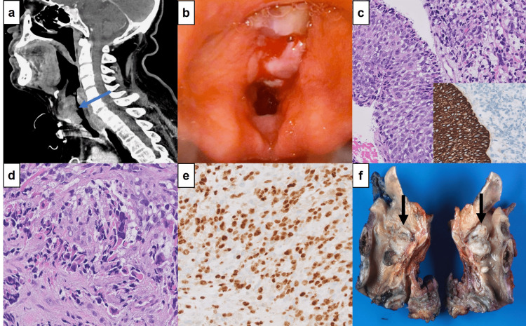
Figure 1. Radiologic and pathologic findings.
(1a) CT neck showing a 4.1 cm heterogeneously enhancing transglottic mass involving supraglottic, glottic, and subglottic regions of the larynx. 1(b) Laryngoscopy showing a large, polypoid mass, involving both left and right true vocal cords and false vocal cords. (1c) Carcinoma in situ adjacent to sarcomatous component (hematoxylin and eosin, x400); CK5/6 showing an immunopositivity in the squamous component but is negative in the sarcomatous component (inset, x200). (1d) High magnification of sarcomatous component showing spindle cells with eosinophilic cytoplasm and enlarged, eccentric, hyperchromatic nuclei with irregular nuclear borders indicative of rhabdomyoblastic differentiation (hematoxylin and eosin, x400). (1e) Myogenin showing diffuse nuclear staining in rhabdomyoblastic component (x200). (1f) Larynx, sagittal section, with large, exophytic glottic mass (arrows), 3.2 cm in greatest dimension.

