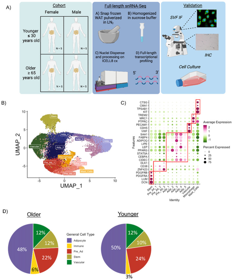
Figure 1. A single nuclei atlas of aging human abdominal subcutaneous white adipose tissue.
Schematic overview of study design and methods (A). UMAP of 25,736 nuclei from abdominal subcutaneous white adipose tissue (WAT) of 10 Younger and 10 Older participants (B). Dotplot of known marker genes for each cell population (C). Cell composition differences in the main cell population between Older and Younger participants (D).

