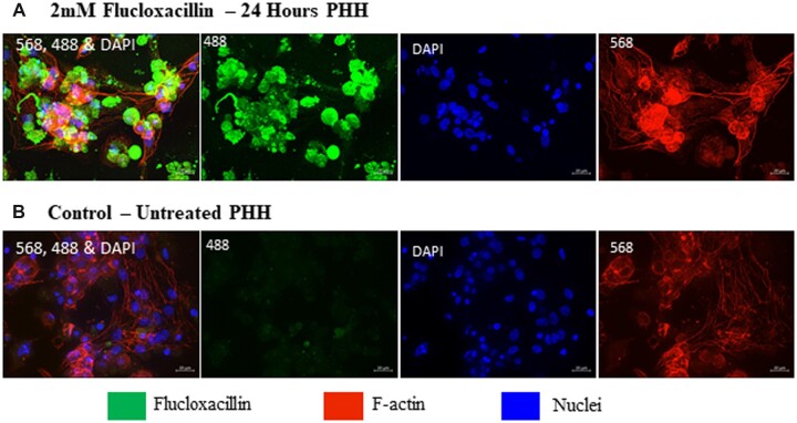
Figure 2.
Qualitative assessment of flucloxacillin binding in PHHs. PHHs were treated with 2 mM flucloxacillin for 24 h to visualize localization and binding of flucloxacillin. Although binding of flucloxacillin (488, green) can be visualized within the cells (A) compared to the untreated control (B), the presence or localization at BC is not clearly defined. 40× magnification, scale bar 20 µm.

