
Fig. 5.
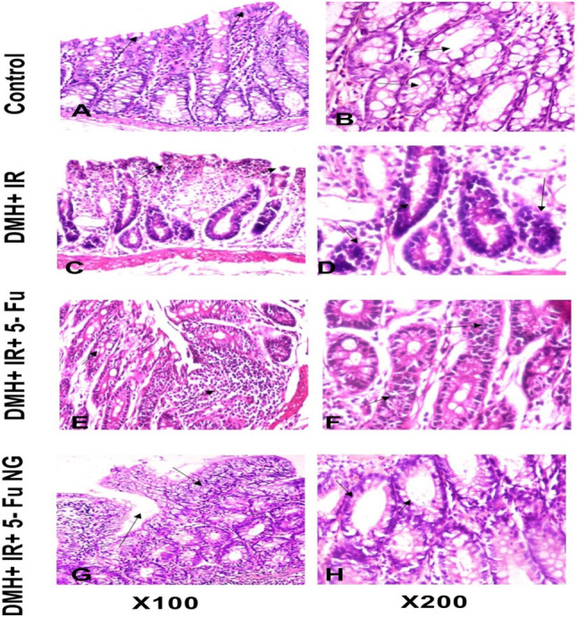
Photomicrograph of the colon tissue section. Figures (A and B) representing control showed a normal histological structure of the colon. However, in Figure (C and D) the DMH + IR showed hyperplasia of the mucosal glands, dysplastic changes, and inflammatory cell infiltration. Figure (E and F) colon tissues after treatment with 5-FU showed moderate dysplasia and some inflammatory cells. Furthermore, 5-FU nanogel lowered the hyperplasia and the inflammatory cells (figure G and H)