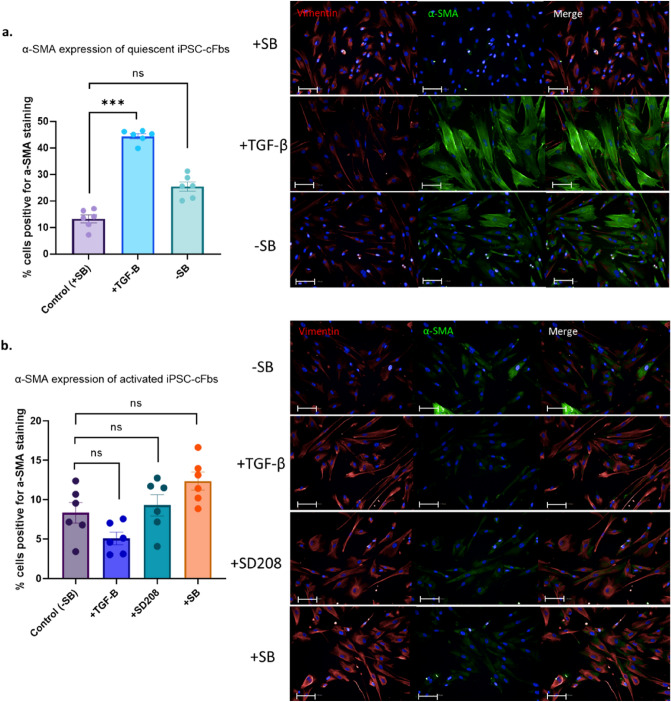
Figure 4.
Immunofluorescence staining of α-SMA in quiescent and activated human induced pluripotent stem cell derived cardiac fibroblasts (hiPSC-cFbs). (a) Quiescent hiPSC-cFbs generated through culture for 2 weeks in the presence of SB435142 were treated with TGF-β. The effect of withdrawal of SB435142 (SB) was assessed. (b) Activated hiPSC-cFbs generated through culture for 2 weeks in the absence of SB435142 were also treated with TGF-β. In addition, the effect of TGF-β receptor inhibition with SD208, and SB435142 (SB) re-introduction was assessed. Data presented as percentage of cells displaying α-SMA expression by immunofluorescence. Scale bar 100 µm. Data presented as mean ± S.E.M. N = 3 plates, 18 wells, nested one-way ANOVA compared with control.

