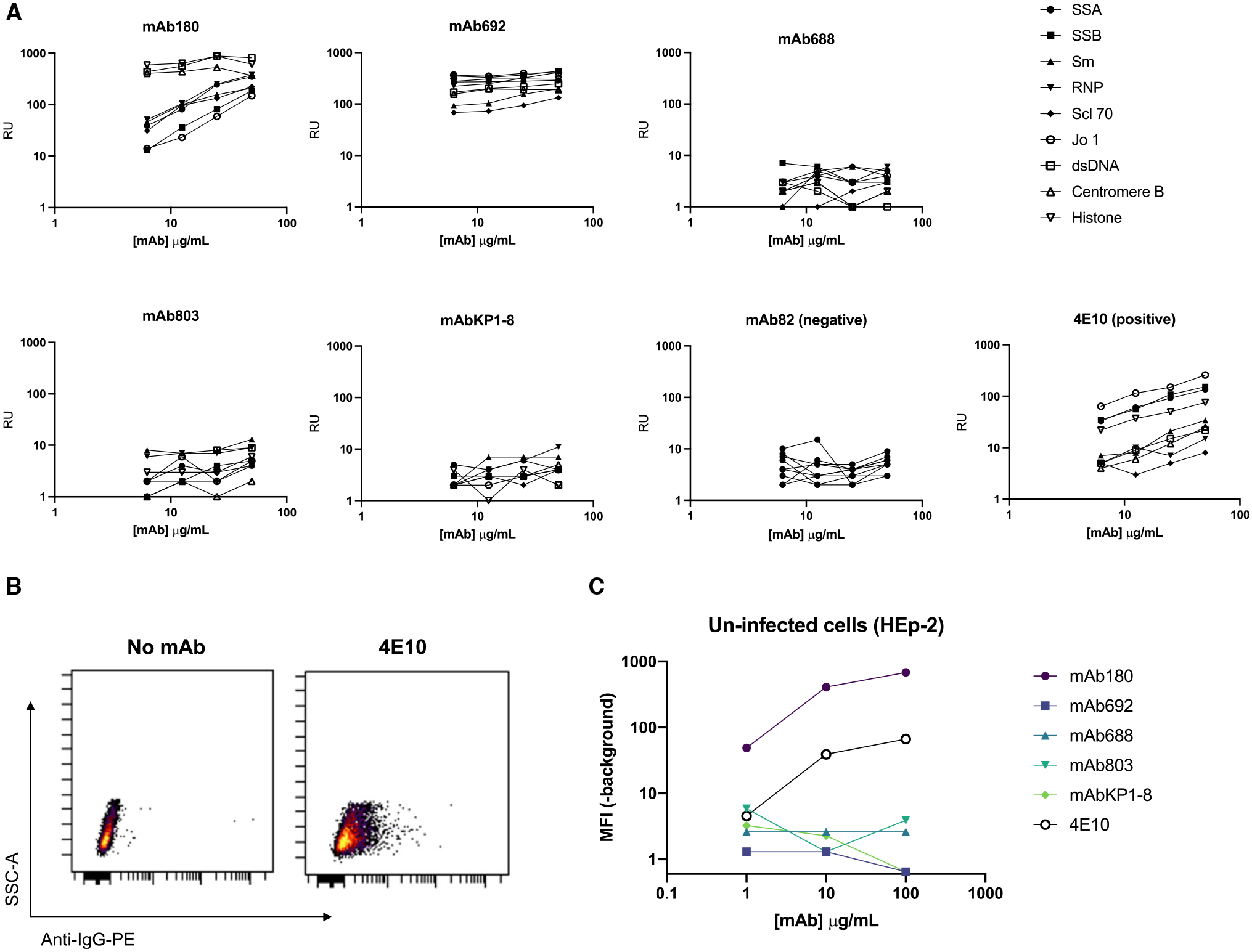
Figure 6. Autoreactivity of HIV-1/HCV cross-reactive antibodies.

(A) Antibody binding to a panel of autoantigens using the AtheNA multiplex assay, over the concentration gradient shown. RUs >100 are considered positive. The HIV-1 antibody 4E10 is shown as a positive control, and mAb82 is shown as a negative control.
(B and C) Antibody binding to whole, unpermeabilized, uninfected HEp-2 cells detected by anti-IgG-PE (anti-IgA-PE; mAb803, mAbKP1–8).
(B) Secondary only (negative) and 4E10 (positive) control plots.
(C) Binding of HIV/HCV cross-reactive antibodies depicted as MFI of the PE channel (-MFI unstained control) at the shown concentration of antibody. AtheNA assays were performed in triplicate. Cell binding experiments were repeated in duplicate.
