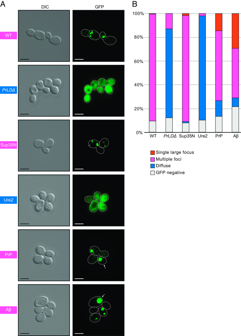
Fig. 4.
Foci detected in cells expressing WT Gag, the GagPrLDΔmutant, and Gag-PrLD chimeras fused to GFP. (A) Live-cell yeast fluorescence microscopy of strains expressing chimeric Gag-GFP after 24-h galactose induction. Differential interference contrast (DIC) and GFP channels are shown with cell outlines added to GFP channels based on DIC images. The strain labels are colored to match the most common foci observed. White arrows indicate cells with a single large focus. Scale bars represent 5 μm. (B) Quantitation of categories of foci observed as a percentage in at least 300 cells. The multiple foci category includes cells with multiple large foci, one or more small foci, or a combination of both sizes. Cell counts are provided in SI Appendix, Table S2.

